Mixed Reality Viewer for Functional Neurosurgery
Visualize
Functional Clinical
Plans in 3D
I’d like a demo
Mixed Reality Viewer for Functional Neurosurgery
Visualize Functional Clinical Plans in 3D
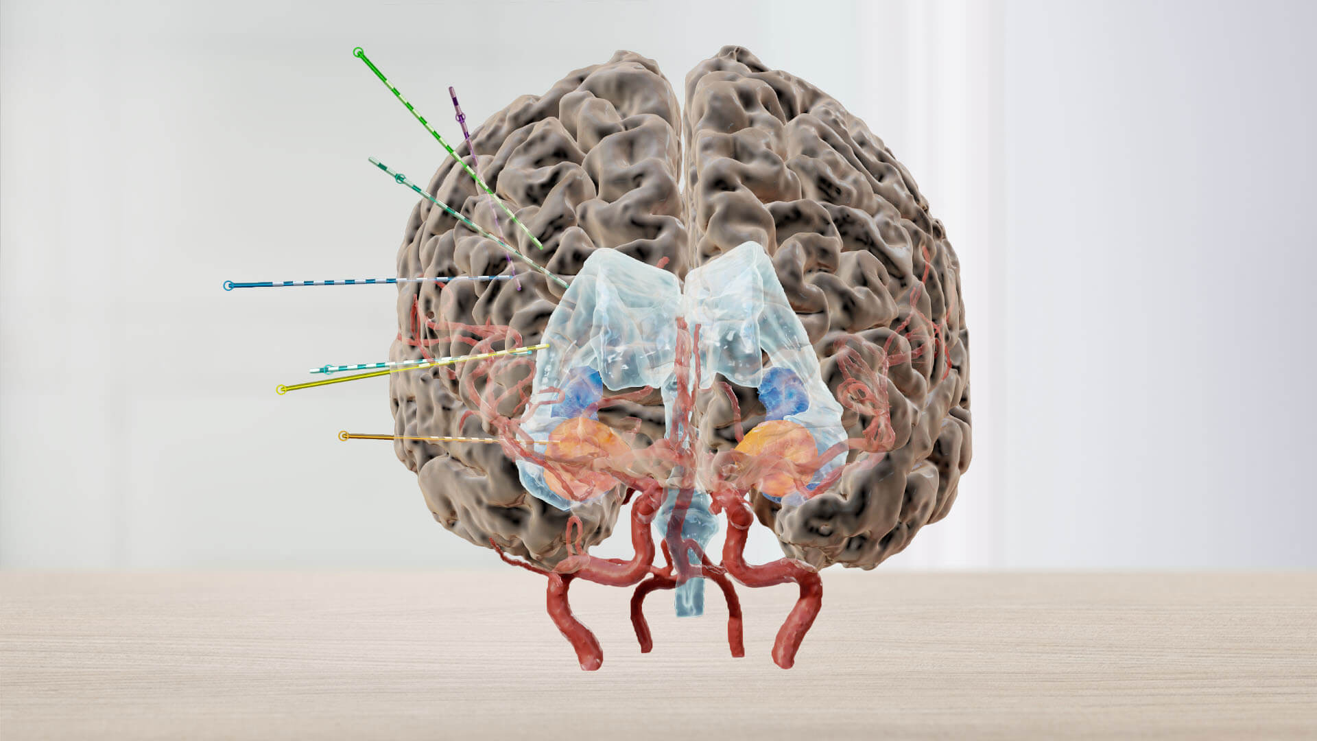
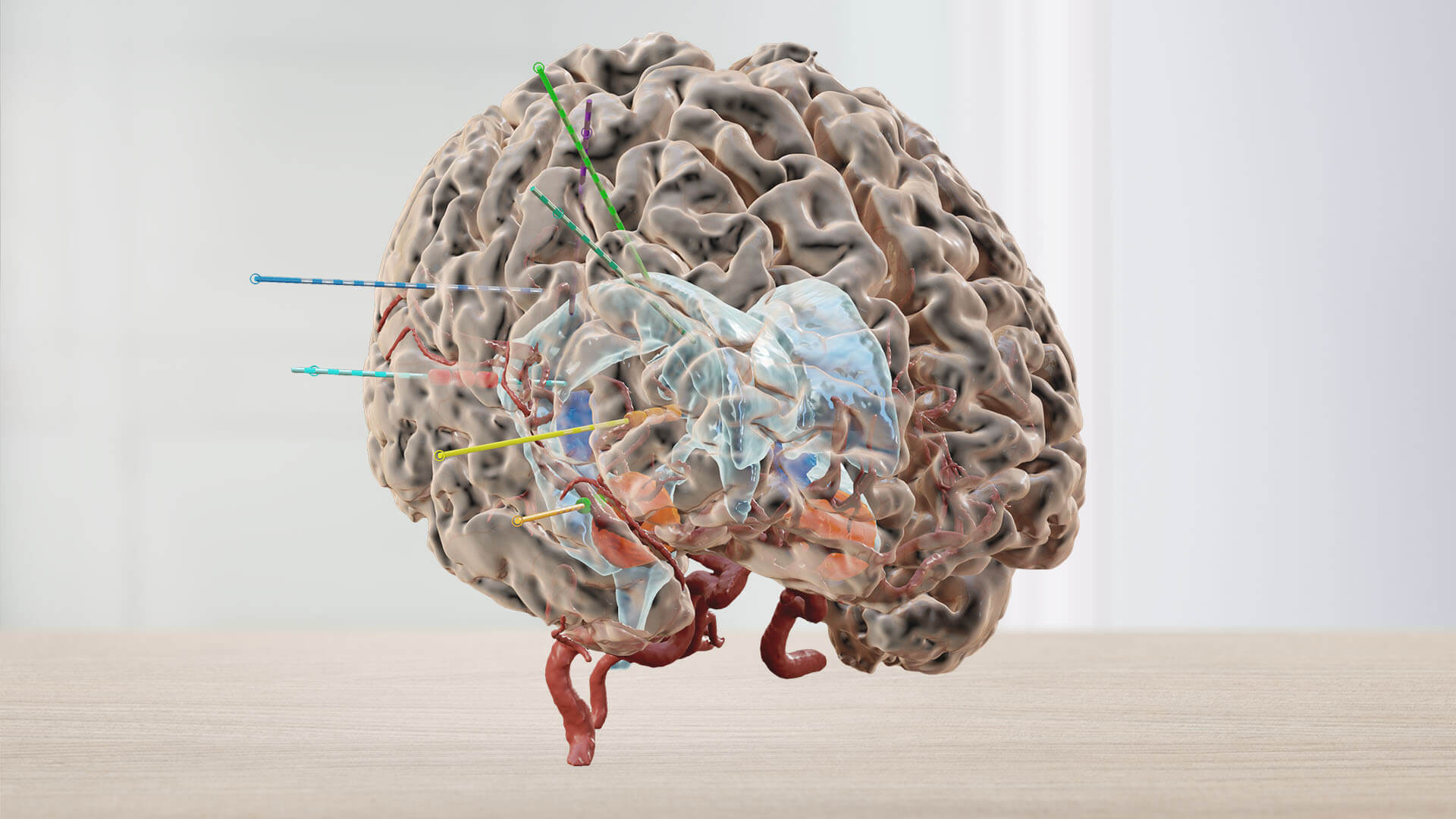
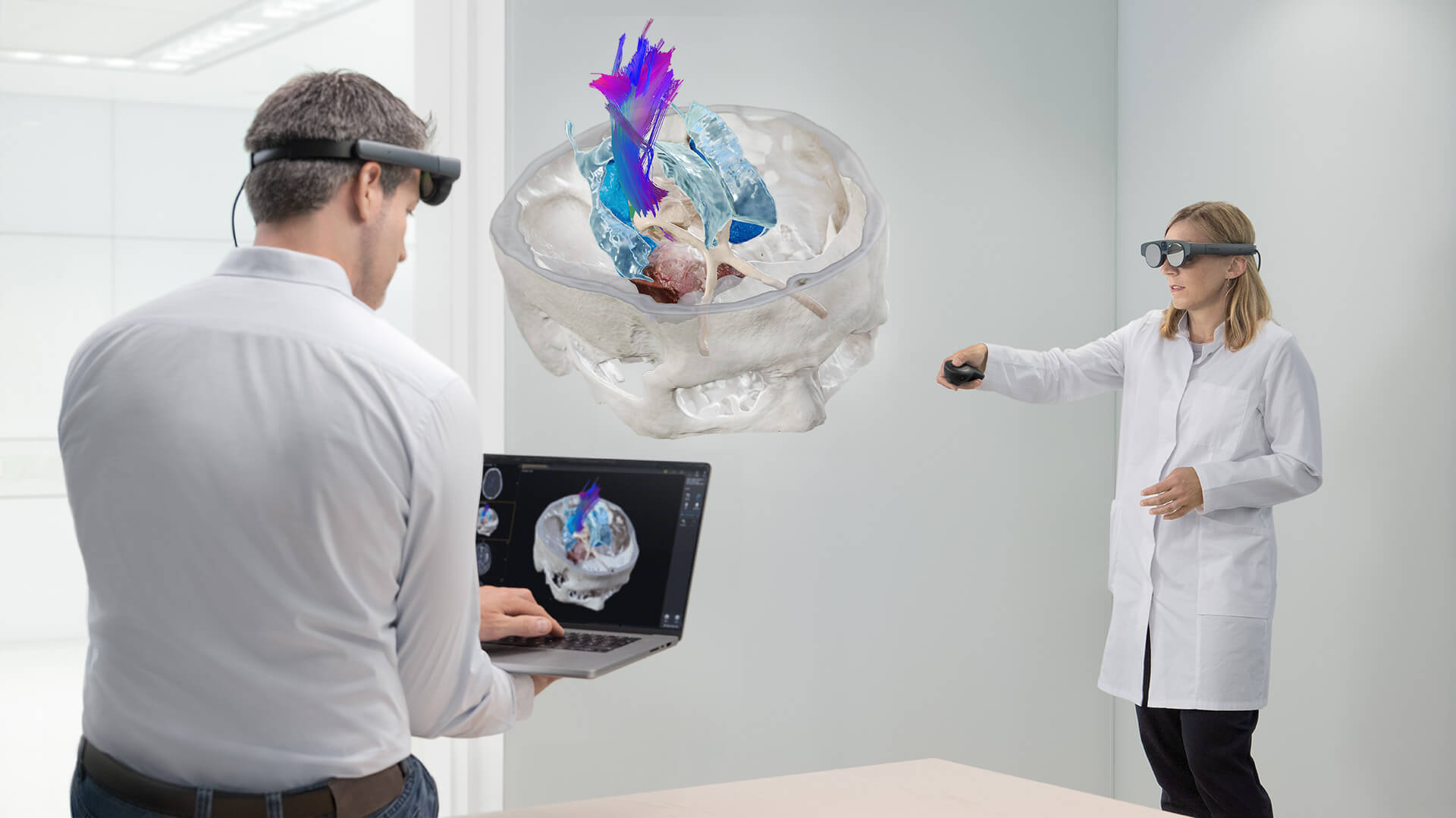
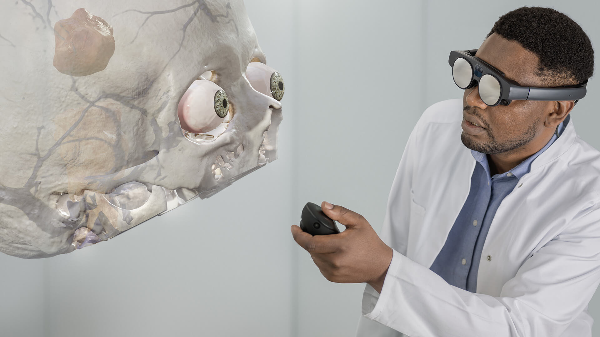
I’d like a demoBring 3D perspectives into functional neurosurgery case review. The Mixed Reality Viewer1 is a technology that produces hyper-realistic 3D patient data. In the context of the Brainlab Functional Neurosurgery portfolio, the Mixed Reality Viewer combined with the Magic Leap 2 provides a 3D visualization of functional clinical plans that include patient-specific anatomical objects, 3D electrode geometry models and white matter tractography.
Accurate 3D visualization for detailed planning review
Accessible & immersive learning opportunities for medical students
Informative patient consultation
Reach out for more.
See how the Mixed Reality Viewer can expand your practice.
Functional Neurosurgery features
in mixed reality
The formulation of plans for invasive diagnostics and treatments, like planning electrodes, has become a multi-disciplinary collaboration. While surgical plans can be prepared with specific Elements applications, mixed reality is an approach that offers an advanced way of viewing the number of electrodes placed and, importantly, understanding the best coverage for the cortex and deeper structures.
Mixed reality for epilepsy
sEEG planning review
- Rotate the patient scans to analyze the cortex for collisions of electrodes.
- Visualize the degree of cortex coverage and major investigative areas (e.g., hippocampus, amygdala).
- Overlay and scroll through 2D data using a trajectory view to minimize vessel collision. Repeat for all potential electrodes.
- Position the patient in a lateral manner to review a single hemisphere.
Mixed reality for epilepsy
sEEG analysis – resection planning review
- Scroll through the relationship of contact location and anatomy during sEEG analysis.
- Utilize the 3D cortex view to hypothesize potential resection.
- Visualize DTI tractography to better understand the relationship between patient anatomy and resection areas.
- Benefit from having a 3D model and data enrichment on a navigation system intraoperatively.
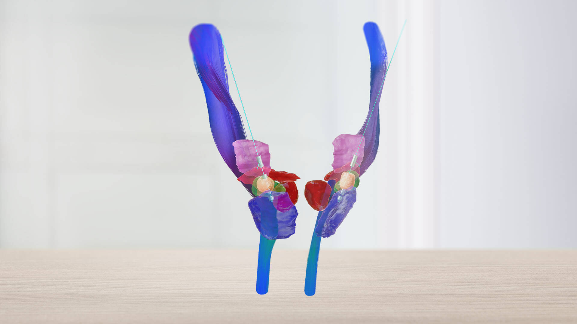
Mixed reality for
deep brain stimulation
- Deep brain stimulation requires the careful planning of implanted leads and the optimization of stimulation parameters. When formulating patient-specific stimulation plans, programming clinicians carefully consider the position of the implanted electrode in the context of the patient’s anatomy in 3D.
- Mixed reality offers a new way to understand and visualize the stimulation field model (SFM) in relation to patient anatomy.
Find your future workflow today
Move patient data from the
screen to reality
With a single click and a glance, your room is digitized for spatial computing. Images are transferred from the Elements Viewer software on screen into the room in front of you with the help of the Magic Leap spatial computing platform.
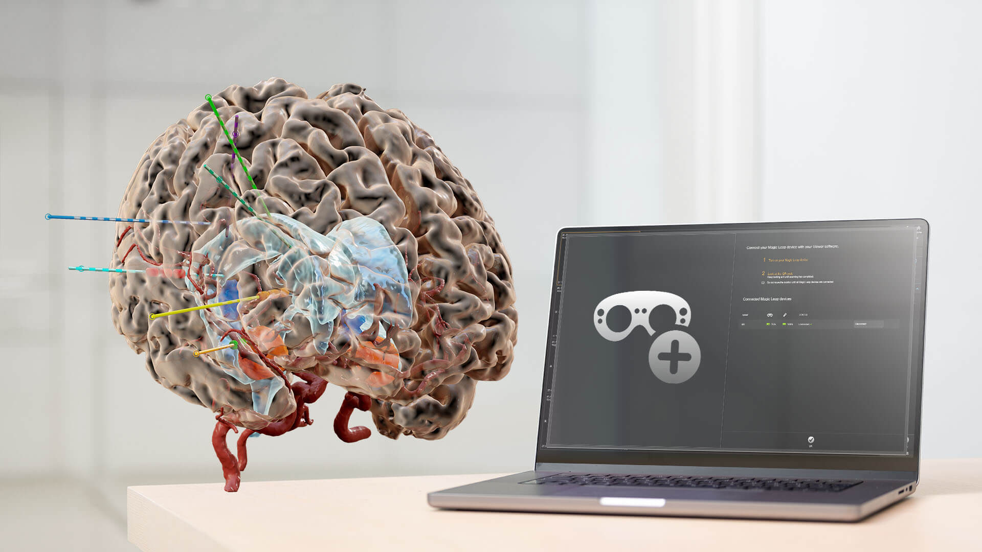
Start with a click
A single click on the Elements Viewer software opens up the world of mixed reality and a new, immersive way to understand patient data.
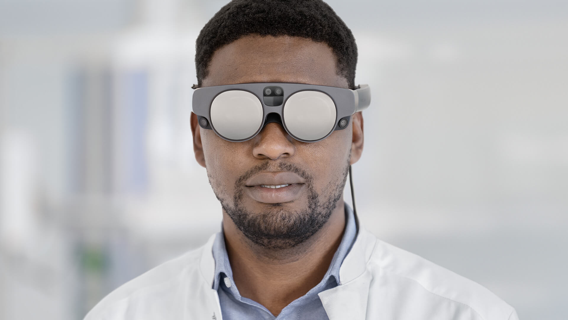
Customizable hardware features
Cameras in the Magic Leap 2 scan and map the room, allowing 3D models and 2D slices to be displayed according to the surgeon’s preference.

Assess, interact, learn
The virtual pointer makes it easy to interact with and highlight the image data and anatomical structures.
Easy integration into unique
clinical workflows
Explore Brainlab mixed reality

1
Not yet commercially available in several countries. Please contact your sales representative.
Bring the best into surgery.
Bring Brainlab.
Contact us today to experience state-of-the-art surgical solutions.